Diabetes Complications: Necrobiosis Lipoidica Diabeticorum
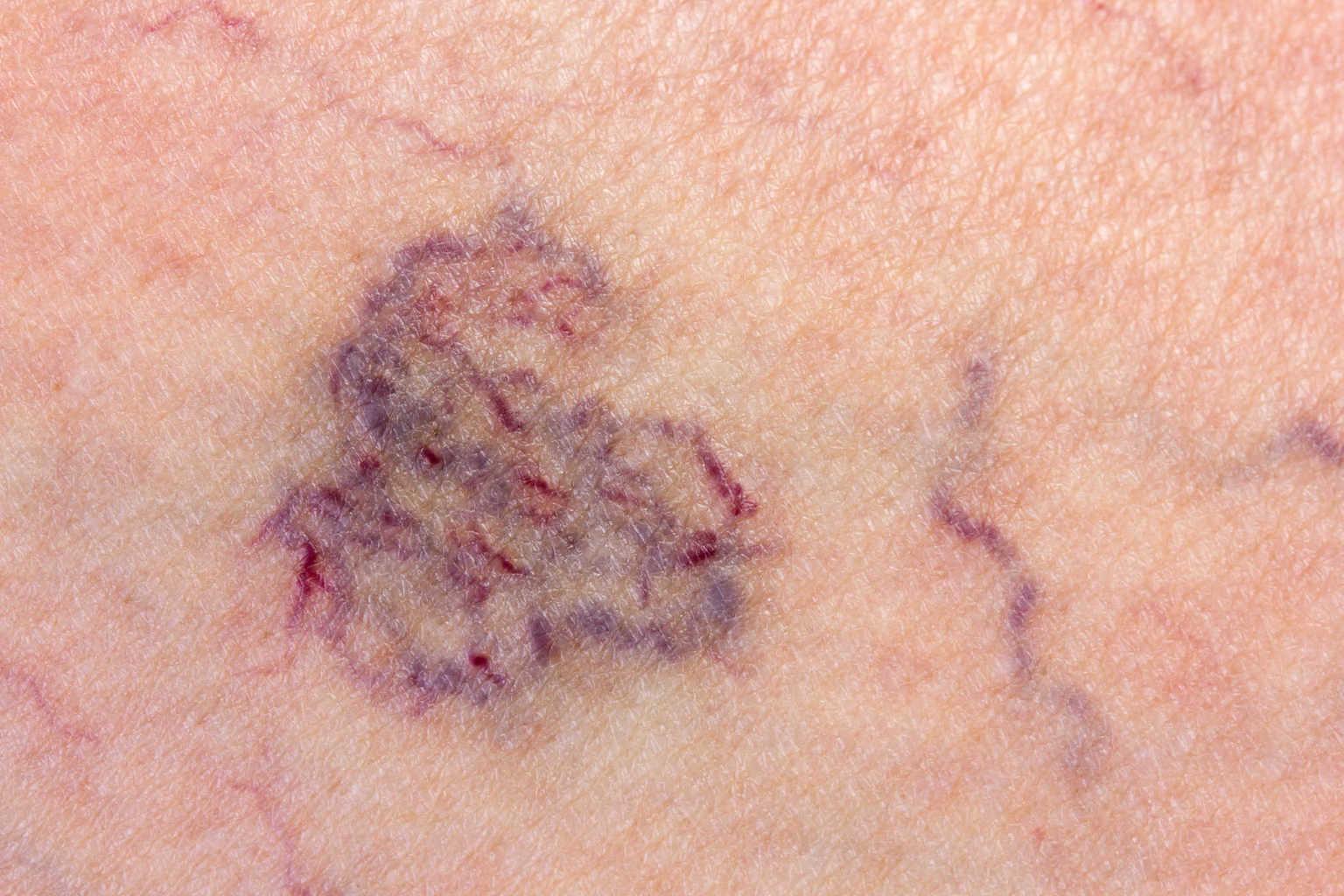
Necrobiosis lipoidica diabeticorum is a degenerative connective tissue disease in the skin. This disease usually appears at the bottom of the foot. The lesions may be small or extend over a large area. The shape is usually prominent, is yellow, and looks waxy, and often has a purple edge.
What is lipoidic necrobiosis?
Necrobiosis lipoidica is a disease characterized by swelling of the skin lesions are calluses with an irregular shape, with reddish-brown pigmentation, and atrophy of the center.
In necrobiosis lipoidica, collagen degenerates with granulomatous response, associated with blood vessels thicken and fat accumulates.
Necrobiosis lipoidica diabeticorum is a rare condition. Adult women are the group most likely to experience it. As long as the wound doesn't open, you don't need to take care of it. However, if you have an open wound, see your doctor for treatment.
Who might experience lipoidic necrobiosis?
Lipoidic necrobiosis is a rare skin condition. Although there is a high prevalence of diabetes mellitus in patients with lipoidic necrobiosis (⅓ cases have diabetes, and ⅔ has glucose tolerance disorders), the prevalence of lipoidic necrobiosis reported in patients with diabetes is 1-2%.
This most often appears in the 30s, but can occur at any age, including babies. This tends to appear earlier in diabetics than others: in one study, about 2% of young people with diabetes (up to 22 years of age) had lipoidic necrobiotic lesions compared with none of the control subjects.
This is 3 times more common in women than in men. Non-diabetic familial groupings of lipoidic necrobiosis do occur, but are very rare.
What are the symptoms of lipoidic necrobiosis?
Necrobiosis lipoidica diabeticorum causes spots similar to diabetic dermopathy, but the numbers are fewer, bigger, and deeper.
Necrobiosis lipoidica diabeticorum often starts with a dull red bump area. After a while, it will look like a shiny scar with a violet-colored edge. Blood vessels under the skin can be seen more easily. Sometimes necrobiosis lipoidica diabeticorum feels itchy and painful. Sometimes the spots break open.
The most common part is the pretibial area, but they can occur on the face, scalp, legs, and upper arms where they tend to be less properly diagnosed. Often there is no pain, but it can also be very painful.
Diabetics with lipoidic necrobiosis appear more frequently in smokers than those who do not smoke, although the presence of lipoidic necrobiosis does not correlate with diabetes control.
How do you manage this condition?
Until now there is no treatment that is truly effective. You must avoid the trauma. Wound care for boils is the same as for other diabetic ulcers. First-line treatment is usually an effective topical steroid. This can reduce inflammation but does not affect burning lesions and can worsen atrophy, so careful monitoring of this case is needed. Intralesional steroid injections also sometimes help, but the risk of ulceration can increase. Your doctor may prescribe the following immunomodulating drugs, with varying degrees of success, to treat lipoidic necrobiosis:
- cyclosporine
- Topical tacrolimus
- Terapi anti-tumor necrosis factor alpha (anti-TNF-α)
Studies show, spontaneous healing of lipoidic necrobiosis after pancreatic and renal transplantation, and immunosuppressive regimes have played an important role in this regard.
In addition, the doctor may give aspirin and dipyridamole. Pentoxifylline reduces blood viscosity and increases fibrinolysis and erythrocyte deformity that might help.
Excision and grafting are sometimes used, but are difficult to heal and often recur.
Treatment may be photodynamic therapy, because it has topical retinoids and topical psoralens with ultraviolet A (PUVA).
Laser treatment has been used to stabilize lesions and reduce erythema and telangiectasias.